Plant Cell Under Electron Microscope - Cell Structure Learning Intention Ppt Video Online Download : Under a high power microscope like the scanning transmission electron microscope, it is.
byNicky Didamo-0
Plant Cell Under Electron Microscope - Cell Structure Learning Intention Ppt Video Online Download : Under a high power microscope like the scanning transmission electron microscope, it is.. She complained that it contained structures showing rough uneven surfaces. Plant cell (under electron microscope). The electron microscope • two types • transmission electron microscope (tem) • scanning electron microscope (sem) • activity • read through the handout on the electron microscope • answer discussion questions ultrastructure of a plant cell as seen through an electron microscope. A cell is a very tiny structure which exists in living bodies. The animal cell is more fluid or elastic or malleable in structure;
Electron microscopes use electron beams focused by electromagnets to magnify and resolve microscopic specimens. The parts of a (palisade) plant cell that can be seen under a light microscope are:cell wallcell (surface) membranelarge (permanent) vacuolecytoplasmnucleuschloroplasts. 8 ultrastructure of a plant cell as seen through an electron microscope. As the wavelength of an electron can be up to 100. Observe an onion cell under the microscope.
Puzzle Of The Day What Is The Link from present5.com Image:plant cell seen under electron microscope. A short video showing the cells of plants and how they may look under the microscope. It is published by the american society of plant biologists. The electron microscope (em) is an impressively powerful microscope that exists today, allowing researchers to view a specimen at nanometer size. Most sem imaging employs the secondary electron detector under high vacuum to provide. Cells of plant or animal tissue. The plant cell as more rigid and stiff. It's a thin slice here is an electron micrograph of an animal cell with the labels superimposed:
However, no obvious structural damage was apparent.
A cell is a very tiny structure which exists in living bodies. Resolving power is the ability to distinguish between separate things which are close to each other. The parts of a (palisade) plant cell that can be seen under a light microscope are:cell wallcell (surface) membranelarge (permanent) vacuolecytoplasmnucleuschloroplasts. What can you see under an electron microscope. Observe an onion cell under the microscope. 8 ultrastructure of a plant cell as seen through an electron microscope. Plant, animal and bacterial cells have smaller components each with a specific function. The detail that can be seen, or resolution, is also important. Light and electron microscopes allow us to see inside cells. For plant cell wall, specimens are generally cut to a thickness of ~80 nm when they are silvery gold in color under. Plant cell under electron microscope. Max knoll at the technical university of munich, he became interested in the possibility of electron microscopy as a. However, no obvious structural damage was apparent.
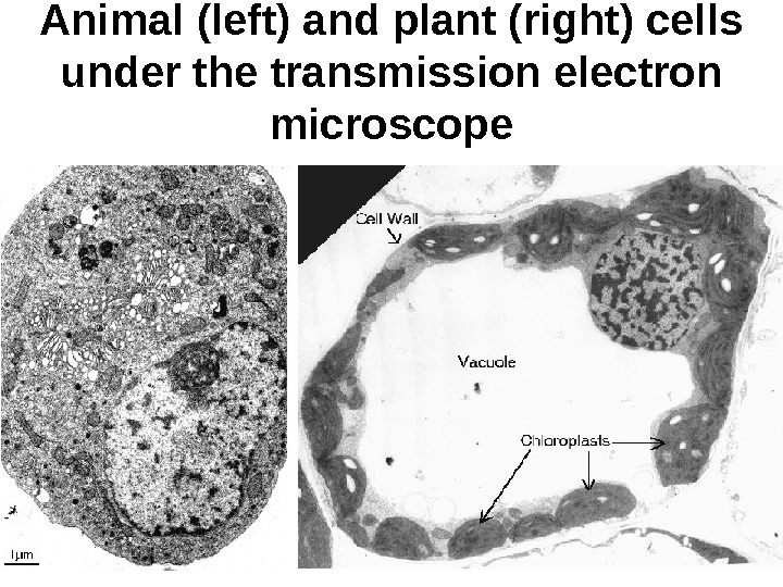
Image:plant cell seen under electron microscope. An electron microscope has a much higher magnifying power and resolution than a regular light microscope. The animal cell is more fluid or elastic or malleable in structure; Image:animal cell seen under electron microscope. Plant cell under the microscope.
Cell Parts Electron Microscope Images Electron Microscope from i.pinimg.com A short video showing the cells of plants and how they may look under the microscope. The detail that can be seen, or resolution, is also important. Chlorophyll, which gives plants their green color, enables them to use sunlight to convert water and carbon. Draw a neat diagram of plant cell and label any three parts which differentiate it from animal cell. Observe an onion cell under the microscope. The electron microscope • two types • transmission electron microscope (tem) • scanning electron microscope (sem) • activity • read through the handout on the electron microscope • answer discussion questions ultrastructure of a plant cell as seen through an electron microscope. Under a high power microscope like the scanning transmission electron microscope, it is. Comparison between a light microscope and an electron microscope:
Under a high power microscope like the scanning transmission electron microscope, it is.
It is published by the american society of plant biologists. 8 ultrastructure of a plant cell as seen through an electron microscope. Ishita observed a slide of eukaryotic cell under electron microscope. Resolving power is the ability to distinguish between separate things which are close to each other. Under a microscope, plant cells are surrounded by cell wall, which are not present in animal cells. Under a high power microscope like the scanning transmission electron microscope, it is. Comparison between a light microscope and an electron microscope: The magnification of a microscope is not the only factor that is important when viewing cells. It's a thin slice here is an electron micrograph of an animal cell with the labels superimposed: However, no obvious structural damage was apparent. Electron microscopes use electron beams focused by electromagnets to magnify and resolve microscopic specimens. An electron microscope has a much higher magnifying power and resolution than a regular light microscope. Under the microscope, animal cells appear different based on the type of the cell.
Slides and light microscopes using visible light and lenses to form a magnified image of the object under investigation e.g. Under the microscope, animal cells appear different based on the type of the cell. Image:animal cell seen under electron microscope. It is published by the american society of plant biologists. It also has a very high resolving power.
Plant Stem Patterns In Nature Microscopic Photography Bio Art from i.pinimg.com Ishita observed a slide of eukaryotic cell under electron microscope. An electron microscope is a microscope that uses a beam of accelerated electrons as a source of illumination. As the wavelength of an electron can be up to 100. What can you see under an electron microscope. Observe an onion cell under the microscope. Here's a photo of a plant cell under an electron microscope. The parts of a (palisade) plant cell that can be seen under a light microscope are:cell wallcell (surface) membranelarge (permanent) vacuolecytoplasmnucleuschloroplasts. It is published by the american society of plant biologists.
Plant cell (under electron microscope).
Plant, animal and bacterial cells have smaller components each with a specific function. Under the microscope, animal cells appear different based on the type of the cell. Image:animal cell seen under electron microscope. An electron microscope has a much higher magnifying power and resolution than a regular light microscope. Max knoll at the technical university of munich, he became interested in the possibility of electron microscopy as a. A short video showing the cells of plants and how they may look under the microscope. For plant cell wall, specimens are generally cut to a thickness of ~80 nm when they are silvery gold in color under. Under a microscope, plant cells are surrounded by cell wall, which are not present in animal cells. 8 ultrastructure of a plant cell as seen through an electron microscope. You see that many features are in common. Cells of plant or animal tissue. It may be hard to believe but the first electron microscope was developed by german physicist hans busch in 1926 with a prototype built by ernst ruska and the electrical engineer max knoll in 1931. Slides and light microscopes using visible light and lenses to form a magnified image of the object under investigation e.g.


Post a Comment